The liver is the largest gland in the body, situated in the right hypochondrium andepigastric region.
Surfaces of the liver
Diaphragmatic surface : smooth, dome-shaped, lies against the inferior surface of the diaphragm.
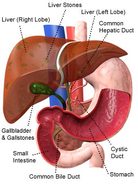
Visceral surface : it is directed inferiorly and is covered with visceral peritoneum except in the fossa of the gallbladder and the porta hepatic.
The surface of the shows :
Gastric impression,
Duodenal impression,
Colic impression,
Right renal impression,
Right suprarenal (adrenal) impression,
Fossae for gallbladder and inferior venacava
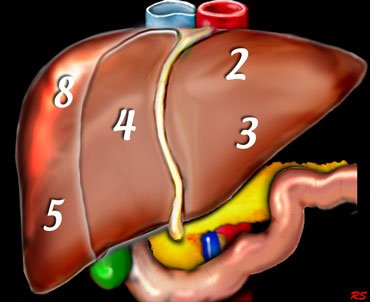
Lobes of the liver:
The right is divided into right and left lobes by fossae for the gallbladder and the inferior vena cava.
The right lobe of the liver is a single large lobe.
The left lobe of the liver is smaller and divided into quadrate and caudate lobes.
Structure of the liver
The liver is covered by Glisson’s capsule.
The basic structural of the liver cell, called hepatocyte. Hepatocytes from layer one or two cells thick, forming anastomosing cords or plates. These plates are separated from each other by large vascular spaces known as liver sinusoids. Hepatic macrophages (kupffer cells) are present among the hepatocytes lining the sinusoids.
Hepatocytes are arranged in hexagonal-shaped lobules. These structural units are called classical liver lobules. The center of each classical lobule is occupied by the central vein.
Where 3 classical lobules contact each other a triangular spaces are demarcated, know as portal lobules with centrally located portal triads. Each portal triad contains a portal venule, a hepatic arteriole and a bile ductile.